The Small Tortoiseshell (Aglais urticae) - Pupation and Hatching
The Small Tortoiseshell (Aglais urticae) is one of the most common butterflies in the family of brush-footed butterflies (Nymphalidae). In Central Europe, the Small Tortoiseshell forms 2 to 3 generations per year. Wintered moths of the last generation can be observed on a sunny day in early spring (Figure 1). The female of the Small Tortoiseshell lays her eggs mainly on the stinging nettles (Urtica dioica). There, the young larvae develop which initially live sociable in webs (Figure 2) and later spread into the environment. Figure 3 shows a mature caterpillar, sunning itself on a nettle leaf.
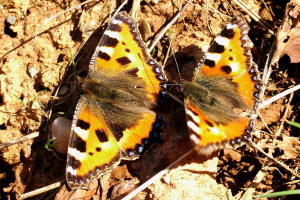 Figure 1: Courtship
 Figure 2: Web with young caterpillars
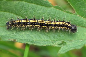 Figure 3: Mature caterpillar


I had found the caterpillar in common with some others on the upper leaves of nettles on 10 May and took three of them. The caterpillars accommodated in a breeding container were agile and always hungry. In the following days they have eaten with great relish some fresh nettle leaves. Once two caterpillars simultaneously marched around on the upper edge of the breeding container. As one caterpillar blocked the way of the other, it was bitten in the buttocks and crashed down. Otherwise, the caterpillars lived together peacefully and focused on the food.
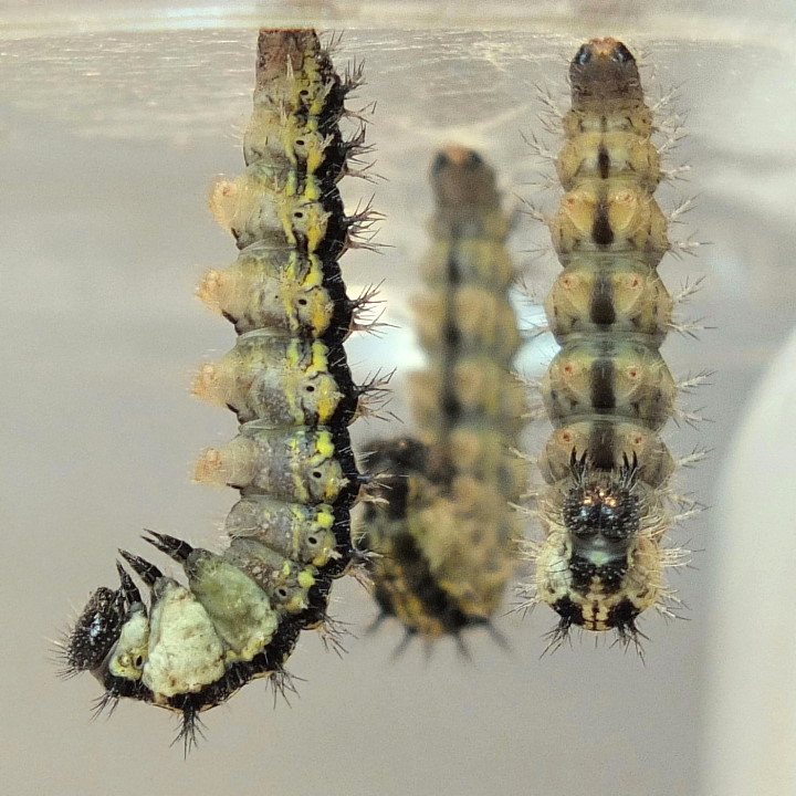
The branches which I had stored in the breeding container as support for pupation were not used by the caterpillars. In the afternoon of May 14 two of three caterpillars were firmly spun on the cover of the breeding container and made first hanging exercises. The 3rd caterpillar was still busy to generate secure attachment. In the evening all caterpillars were hanging on the cover head-down. Since they were not pupated in the morning, I carefully removed the cover and built a temporary construction for picture taking. Figure 4 shows the three caterpillars about 100 min before pupation of the first caterpillar.
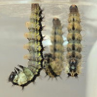 Figure 4: 100 min before pupation of the first caterpillar.
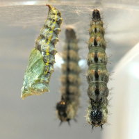 Figure 5: The first caterpillar pupated and threw larval skin away.

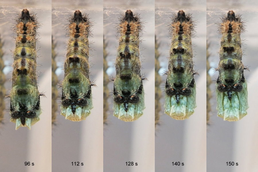
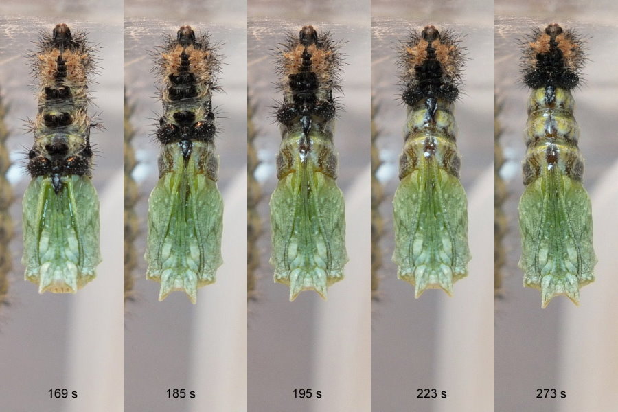
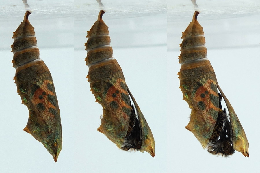
Despite frequent controls, I missed the beginning of pupation of the first caterpillar. When I looked up again, I found already a pupa (Figure 5) which made some rocking movements to get rid of the stripped caterpillar skin. In the hope that the second caterpillar follows the first one, I aligned the camera to it and waited. Fortunately, the second caterpillar started almost immediately with pupation, which is shown in the following Figures 6 - 8. Unfortunately, the lighting conditions were very bad, because I could not use a flash to make sure that the caterpillar does not feel disturbed by the flash light. The first, or rather, last image of the second caterpillar was created a few minutes before pupation. Therefore, the time scale starts on Figure 6 in the second field. After about 5 minutes, the second caterpillar was pupated. I find the rapid change in physiques from the caterpillar to the significantly shorter and wider pupa remarkable, which was completed already in the first 2 minutes. A few hours later the third caterpillar had pupated. The event I missed again.
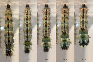 Figure 6: The second caterpillar starts pupation,
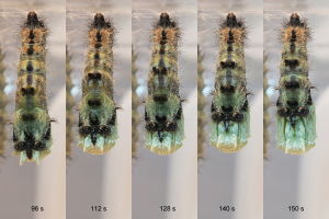 Figure 7: sheds its skin
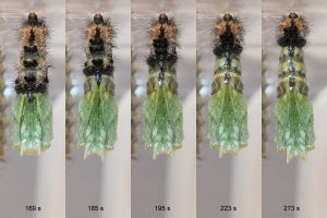 Figure 8: and presents new soft green outfit.

After almost 12 days pupal stage the first two butterflies were hatched (Figure 9) in the early morning of 27 May. They had already unfolded their wings and hung on the empty puparia. In order to observe the slip of the third butterfly, I gently transported the cover with the first butterflies and the pupa to the balcony and put it on a camera-oriented position. After a recovery period for hardening the wings the first two Small Tortoiseshells flew in time to freedom before the slip of the third butterfly. Figure 10 shows the first butterflies in preparation for launch, which took place at 9:40 clock.
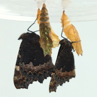 Figure 9: The first two butterflies are hatched.
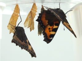 Figure 10: Immediately before take-off


When comparing the fresh pupa of Figure 5 with the pupa from the first field of Figure 11 it becomes immediately apparent that the color of the pupa has changed during the pupal period. The fresh pupa is pale green and still shows the yellow stripes of the caterpillar. These have disappeared during the pupal period. Just before hatching the conspicuous wing pattern of the Small Tortoiseshell shimmers through the pupal case (Figure 11).
Last year I had reared a Small Tortoiseshell. This butterfly also did not use the branches offered for pupation and had hung on the cover of the breeding container. Approximately one-half to one hour before the slip a loud rattling was heard on the window sill. When I was looking for the cause, I saw that the pupa rocked heavily. The rattling noise was generated by the collision of the pupa with the container lid. Since the activities were repeated several times with short pauses in between, I used the opportunity to record a video with the mobile phone.
This strange behavior of the pupa observed in the past year was the reason to pick up caterpillars this year again. The last pupa of this year, which was attached to the lid just like the pupa from the previous year, did not show this hyperactive behavior and only moved slightly just before hatching.
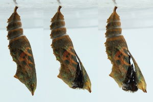 Figure 11: The puparium bursts.
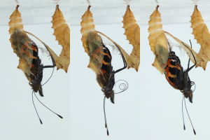 Figure 12: The 3rd butterfly extracts leggs, antennae and proboscis,
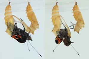 Figure 13: emerges completely, clings to the puparium
 Figure 14: and starts to unfold its wings.
 Figure 15: The wings grow
 Figure 16: and must harden. Now the Small Tortoiseshell is ready for take-off.
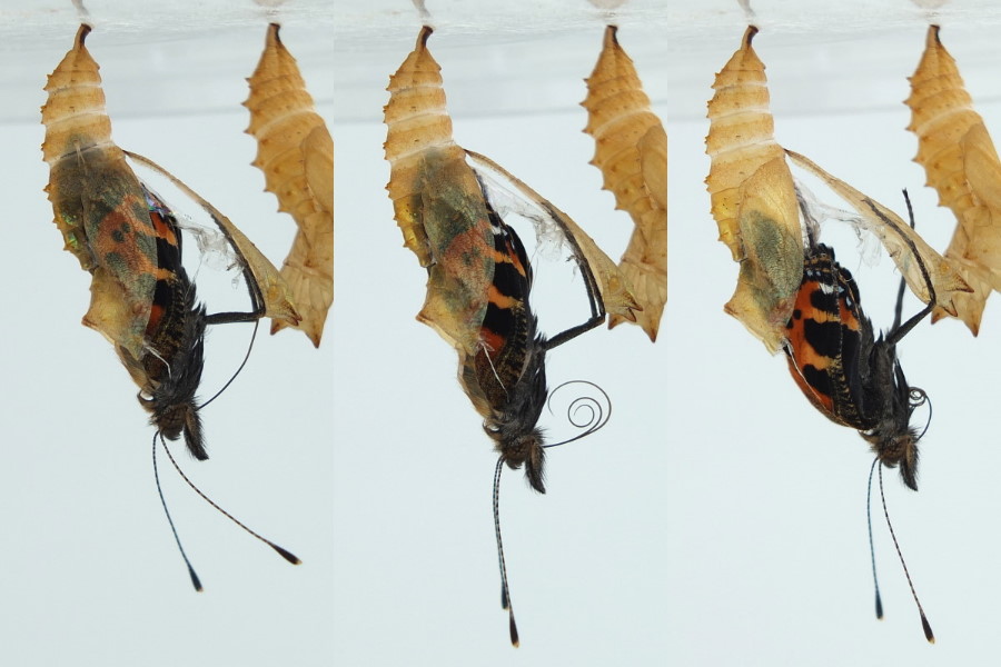

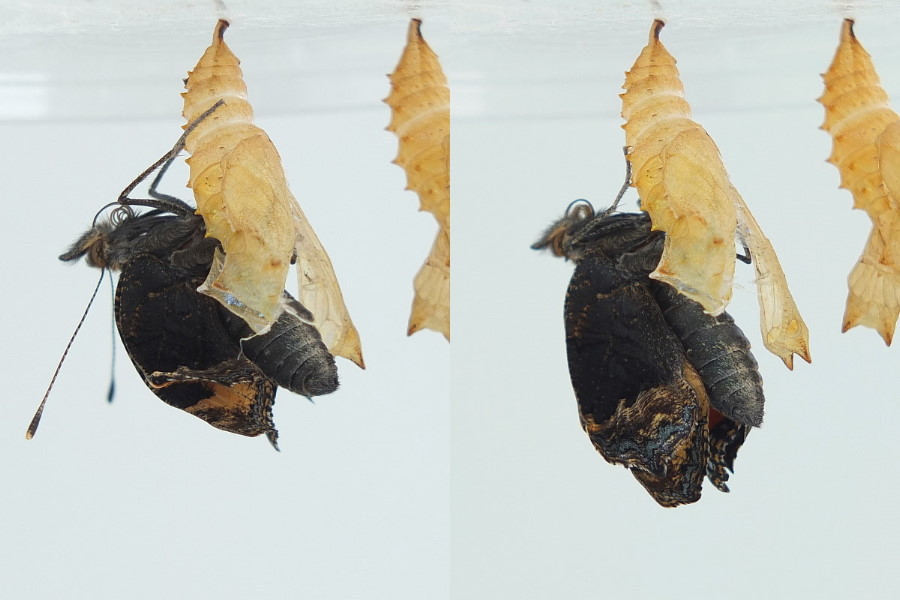


At 10.30 a.m. the puparium bursted and the third butterfly began to slip out gently. As can be seen in Figure 12 its freshly curled proboscis consists of 2 parts. The butterfly completely slipped out of the pupal case, made a skillful turn and clung to the puparium (Figure 13). The Figures 14 and 15 show the butterfly while inflating the wings. For that, hemolymph is pumped into the veins. After inflation the soft wings have to harden for a while (Figure 16). Overall, the inflation and hardening of the wings took about two and a half hours. Shortly after 1 p.m. the last Small Tortoiseshell has departed.